Growth hormones are not FGF23, but share some Vitamin D gene interactions
Yes and no — they're unrelated molecules, but they're not unconnected biologically.
Different in almost every structural sense:
| Growth hormone | FGF23 | |
|---|---|---|
| Source | Pituitary somatotrophs | Osteocytes and osteoblasts |
| Gene family | Somatotropin/prolactin | Fibroblast growth factor (22 members) |
| Receptor | GHR → JAK2/STAT5 | FGFR1c, but only with α-Klotho as co-receptor |
| Main job | Longitudinal growth, lipolysis, IGF-1 release | Phosphate excretion and shutting down calcitriol |
Part of the confusion is naming. "Fibroblast growth factor" is a historical label from the first family members, which were mitogenic for fibroblasts in culture. FGF23 is one of the three "endocrine" FGFs (with FGF19 and FGF21) that escaped that role entirely — it circulates as a hormone and has essentially nothing to do with fibroblast proliferation. Meanwhile GH's actual effector, IGF-1, is a growth factor. So the two categories overlap in vocabulary but not in lineage.
Where they do intersect:
The clearest link is phosphate handling, where they pull in opposite directions. GH/IGF-1 increases proximal tubule phosphate reabsorption and stimulates CYP27B1, raising calcitriol. FGF23 downregulates NaPi-2a/2c, suppresses CYP27B1, and induces CYP24A1 — lowering calcitriol. Functional antagonists on the same target tissue.
They also both matter at the growth plate. In X-linked hypophosphatemic rickets, chronically high FGF23 causes short stature, and recombinant GH has been used as an adjunct (with mixed results, since it can worsen the phosphate wasting).
The FGF23 side is the one that keeps showing up: it's the brake on the vitamin D system. Calcitriol induces FGF23 transcription, and FGF23 then suppresses calcitriol production — a closed feedback loop that partly explains why serum 1,25(OH)₂D doesn't climb linearly with intake, and why FGF23 rises in CKD long before phosphate does.
How FGF23 regulates the vitamin D metabolism
FGF23 sits at the top of the phosphate arm of mineral regulation, and vitamin D is one of its main levers.
The signal
Osteocytes and osteoblasts secrete FGF23 in response to phosphate load, 1,25(OH)₂D, PTH, iron deficiency, inflammation (IL-6, IL-1β), and erythropoietin. It reaches the kidney and signals through FGFR1c (also 3c/4) — but only where α-Klotho is present as an obligate co-receptor. Klotho is what makes the proximal and distal tubule the primary targets rather than every FGFR-bearing tissue. Downstream it runs through the RAS–ERK1/2–MAPK cascade to EGR1.
The double hit on calcitriol
- Suppresses CYP27B1. Transcription of 1α-hydroxylase in the proximal tubule falls within hours. Less 25(OH)D → 1,25(OH)₂D conversion.
- Induces CYP24A1. 24-hydroxylase rises, accelerating catabolism of 1,25(OH)₂D to 1,24,25 and onward to calcitroic acid.
Both arms push the same direction, which is why the calcitriol drop after an FGF23 rise is fast and steep compared with anything PTH does. The second arm matters more than it usually gets credit for: CYP24A1 also 24-hydroxylates 25(OH)D itself to 24,25(OH)₂D. So sustained FGF23 elevation doesn't only lower the active hormone — it shortens the half-life of the substrate pool, which is a plausible contributor to the "needs an unusually large dose to move the needle" phenotype.
The feedback loop closes
1,25(OH)₂D binds VDR–RXR at VDREs in the FGF23 promoter and drives osteocyte FGF23 transcription. So calcitriol induces its own suppressor. This is the loop that makes calcitriol and active analogs in CKD raise FGF23 substantially, while cholecalciferol at physiological doses generally moves FGF23 little — plain D₃ raises 25(OH)D without proportionally raising 1,25(OH)₂D, because renal CYP27B1 is under its own tight control. Bolus dosing is the exception where the induction becomes measurable.
FGF23 also suppresses PTH (Klotho-dependent, in parathyroid) and downregulates NaPi-2a/2c to produce phosphaturia — so the calcitriol suppression and the reduced gut absorption of phosphate (via less 1,25(OH)₂D acting on NaPi-2b) work together to unload phosphate.
Where this shows up clinically
Any high-FGF23 state gives you low 1,25(OH)₂D with a normal-looking 25(OH)D: XLH, ADHR, tumor-induced osteomalacia, fibrous dysplasia, and above all CKD — where FGF23 rises very early, well before serum phosphate becomes abnormal, and is arguably the initiating lesion in CKD-MBD rather than a consequence.
Two modifiers worth keeping in view for your phosphate-additive work:
- Klotho falls with age and with CKD. Declining Klotho creates tissue-level FGF23 resistance, which drives FGF23 higher still, and at very high concentrations FGF23 acts on FGFR4 in cardiomyocytes without Klotho — the off-target LVH pathway. So the same serum FGF23 does not mean the same renal effect in an 80-year-old as in a 30-year-old.
- Iron status. Iron deficiency raises FGF23 transcription and, in ADHR, blocks cleavage of intact FGF23 — a cheap, correctable input to the whole loop that rarely gets checked alongside 25(OH)D.
The interstitial angle you've been working is consistent here: FGF23's suppression of renal CYP27B1 constrains the endocrine 1,25(OH)₂D pool, but extrarenal CYP27B1 is substrate-limited by 25(OH)D delivery, not by FGF23 — except insofar as CYP24A1 induction erodes that substrate. That distinction is probably the most useful thing to keep separate when writing this up.
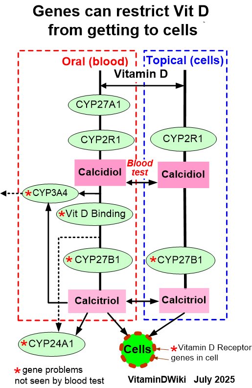
Some Vitamin D Genes are not noticed by a standard Vitamin D test