C15:0 (Fatty15, Pentadecanoic acid) a fatty acid from grass-fed animals may be essential - several studies
C-15:0 may be an essential fatty acid - review Dec 2024
New insights on pentadecanoic acid with special focus on its controversial essentiality: A mini-review
Biochimie Volume 227, Part B, Dec 2024, Pages 123-129 https://doi.org/10.1016/j.biochi.2024.10.008

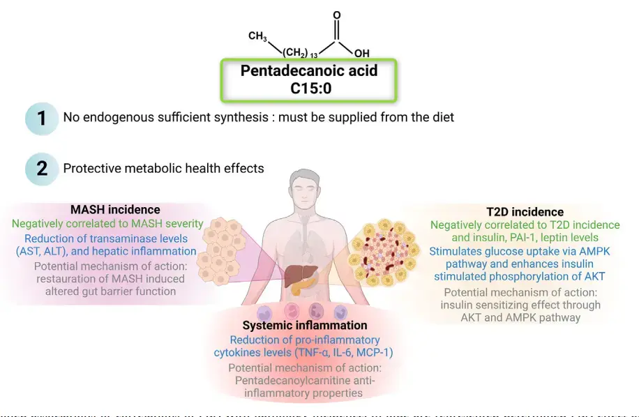
Pentadecanoic acid (C15:0, PDA ) is an odd and minor fatty acid that has been neglected in the literature until the last decade. Indeed, as a specific fatty acid of dairy fat, PDA was only used as a biomarker of dairy fat consumption. Lately, PDA was first correlated negatively with the incidence of metabolic syndrome disorder, then its physiological effects have been investigated as a protective fatty acid. PDA supplementation has been demonstrated as negatively correlated with elevated levels of leptin, plasminogen activator inhibitor-1 and insulin, and has been shown to exhibit sensitizing insulin effects with activation of AMPK pathway. PDA also reduced the severity of metabolic dysfunction-associated steatohepatitis (MASH), notably through reduced alanine transaminase and pro-inflammatory cytokines levels. The final effect described for PDA is its ability to display anti-inflammatory properties in several pathology models. Hence, considering these multiple effects, the presence of PDA could be associated with a healthier physiological state, this raises the question of whether the presence of PDA in the body, in adequate quantities, is needed to participate to health maintenance. PDA is not synthesized in sufficient quantities endogenously, so it must be provided by the diet, mainly through dairy fat, although other types of food can also contribute to the dietary intake of PDA . Essential fatty acids are described as not being endogenously synthesized in sufficient and required quantities to maintain physiological health.
Thus, PDA might gather both conditions to be described as essential, yet further investigations on both criteria are needed to enhance knowledge on this odd chain fatty acid with promising impact as potential protective supplement nutrient.
📄 Download the PDF from VitaminDWiki
C15:0 — Found in Dairy — May Be an Essential Fat - Mercola Aug 2024
Includes a 1 hour video by Venn-Watson
" C15:0, also known as pentadecanoic acid , is an odd-chain saturated fat primarily found in dairy products, some fish, and certain plants. The story of C15:0's importance begins in an unlikely place — with dolphins."
"Dr. Stephanie Venn-Watson , a veterinary epidemiologist, was brought on board the Navy's Marine Mammal program about 20 years ago to help understand aging in dolphins and protect their health. The Navy has been caring for a population of dolphins for over 60 years, and these dolphins are living much longer in captivity (40 to 50+ years) compared to their wild counterparts (around 20 years)."
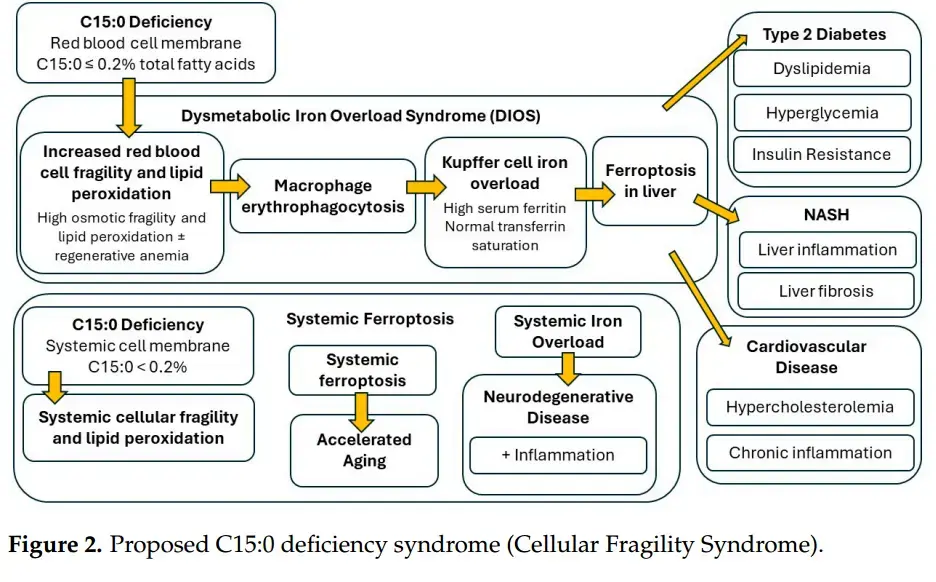
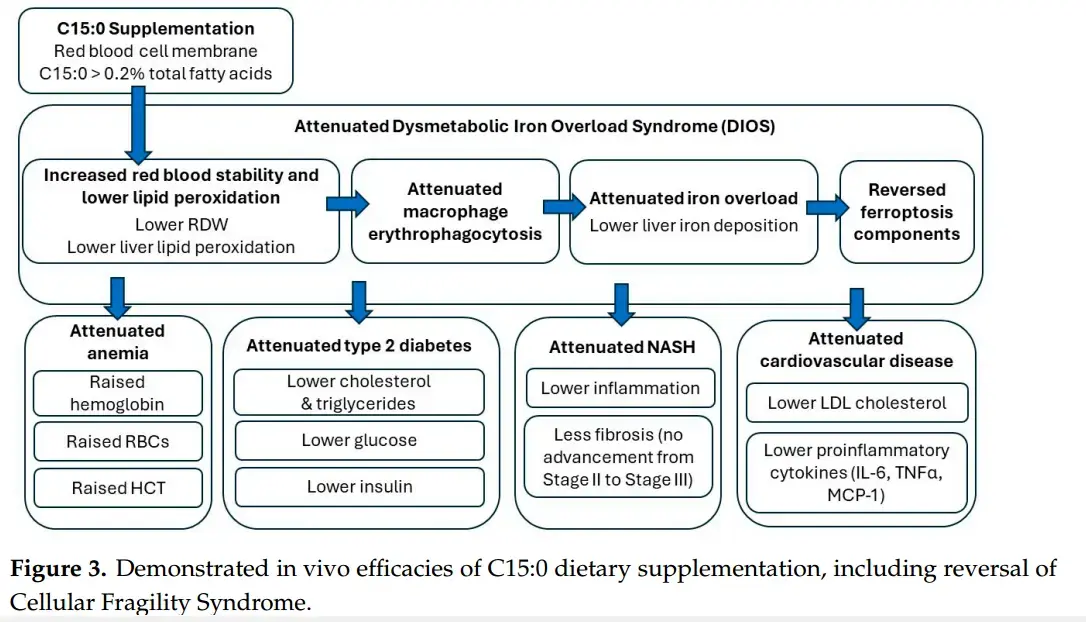
"Dr. Venn-Watson's research suggests that when your C15:0 levels drop below 0.2% of total fatty acids in your cell membranes, you enter a state she calls "Cellular Fragility Syndrome." This syndrome is characterized by fragile red blood cells, anemia, iron overload in the liver, and increased risk of conditions like Type 2 diabetes, cardiovascular disease and fatty liver disease. It's a domino effect that starts at the cellular level and cascades into systemic health issues."
"The impact of C15:0 deficiency is far-reaching and multifaceted. The "Cellular Fragility Syndrome" resulting from C15:0 deficiency is characterized by a cascade of health issues. It starts with fragile red blood cells susceptible to lipid peroxidation, leading to anemia and dysmetabolic iron overload syndrome (DIOS). This iron overload can trigger ferroptosis in the liver, potentially leading to advanced nonalcoholic fatty liver disease (NAFLD) and nonalcoholic steatohepatitis (NASH)."
"The syndrome also encompasses insulin resistance, metabolic syndrome, Type 2 diabetes, and cardiovascular disease. Perhaps most alarmingly, it includes systemic iron overload and ferroptosis, which can accelerate aging and tissue damage throughout your body. It's a complex web of interconnected health issues, all potentially stemming from a deficiency in this one crucial fat."
"Based on the available research, your cell membranes require more than 0.2% C15:0 to ensure cellular stability. Optimal circulating C15:0 concentrations should be between 0.4% to 0.64% of total fatty acids. C15:0 deficiency is defined as 0.21% or less of total circulating fatty acids"
The Cellular Stability Hypothesis: Evidence of Ferroptosis and Accelerated Aging-Associated Diseases as Newly Identified Nutritional Pentadecanoic Acid (C15:0) Deficiency Syndrome - June 2024
Metabolites 2024, 14(7), 355; https://doi.org/10.3390/metabo14070355
by Stephanie Venn-Watson 1,2ORCID
1 Seraphina Therapeutics Inc., San Diego, CA 92106, USA
2 Epitracker Inc., San Diego, CA 92106, USA


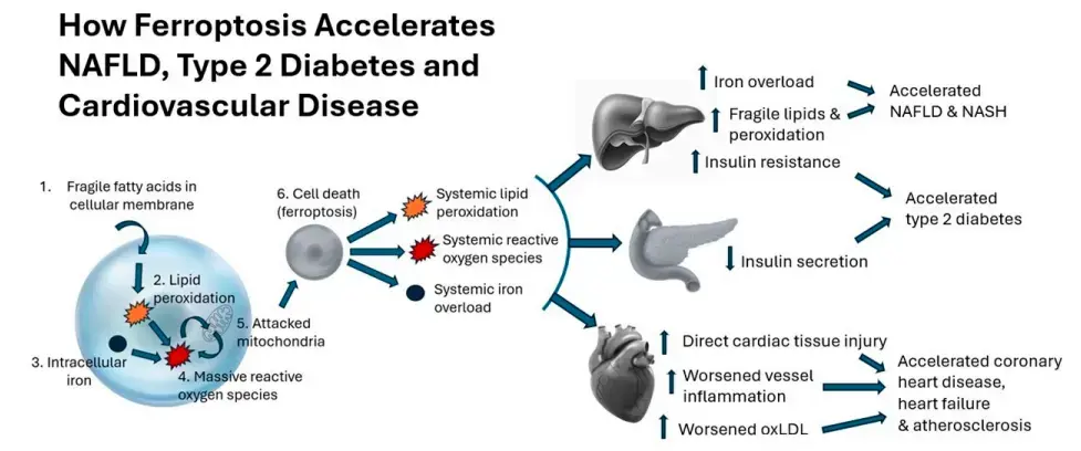
Ferroptosis is a newly discovered form of cell death caused by the peroxidation of fragile fatty acids in cell membranes, which combines with iron to increase reactive oxygen species and disable mitochondria. Ferroptosis has been linked to aging-related conditions, including type 2 diabetes, cardiovascular disease, and nonalcoholic fatty liver disease (NAFLD). Pentadecanoic acid (C15:0), an odd-chain saturated fat, is an essential fatty acid with the primary roles of stabilizing cell membranes and repairing mitochondrial function. By doing so, C15:0 reverses the underpinnings of ferroptosis.
Under the proposed “Cellular Stability Hypothesis”, evidence is provided to show that cell membranes optimally need >0.4% to 0.64% C15:0 to support long-term health and longevity. A pathophysiology of a newly identified nutritional C15:0 deficiency syndrome (“Cellular Fragility Syndrome”) is provided that demonstrates how C15:0 deficiencies (≤0.2% total circulating fatty acids) can increase susceptibilities to ferroptosis, dysmetabolic iron overload syndrome, type 2 diabetes, cardiovascular disease, and NAFLD. Further, evidence is provided that C15:0 supplementation can reverse the described C15:0 deficiency syndrome, including the key components of ferroptosis. Given the declining dietary intake of C15:0, especially among younger generations, there is a need for extensive studies to understand the potential breadth of Cellular Fragility Syndrome across populations.
📄 Download the PDF from VitaminDWiki
References
Hulbert, A.J. On the importance of fatty acid composition of membranes for aging. J. Theor. Biol. 2005,234, 277-288. [CrossRef] [PubMed]
Dixon, S.J.; Lemberg, K.M.; Lamprecht, M.R.; Skouta, R.; Zaitsev, E.M.; Gleason, C.E.; Patel, D.N.; Bauer, A.J.; Cantley, A.M.; Yang, W.S.; et al. ferroptosis : An iron-dependent form of nonapoptotic cell death. Cell 2012,149,1060-1072. [CrossRef]
Miao, R.; Fang, X.; Zhang, Y.; Wei, J.; Zhang, Y.; Tian, J. Iron metabolism and ferroptosis in type 2 diabetes mellitus and complications: Mechanisms and therapeutic opportunities. Cell Death Dis. 2023,14,186. [CrossRef] [PubMed]
Zhang, H.; Zhang, E.; Hu, H. Role of ferroptosis in non-alcoholic fatty liver disease and its implications for therapeutic strategies. Biomedicines 2021, 9,1660. [CrossRef] [PubMed]
Pasini, A.M.F.; Stranieri, C.; Busti, F.; Di Leo, E.G.; Girelli, D.; Cominacini, L. New insights into the role of ferroptosis in cardiovascular disease. Cells 2023,12, 867. [CrossRef] [PubMed]
Xu, Y.; Zhao, Y.; Zhou, L.; Qiao, H.; Xu, Q.; Liu, Y. The role of ferroptosis in neurodegenerative diseases. Mol. Biol. Rep. 2023, 50, 1655-1661. [CrossRef] [PubMed]
Tonnies, T.; Brinks, R.; Isom, S.; Dabelea, D.; Divers, J.; Mayer-Davis, E.J.; Lawrence, J.M.; Pihoker, C.; Dolan, L.; Liese, A.D.; et al. Projections of type 2 and type 2 diabetes burden in the U.S. population aged < 20 years through 2060: The SEARCH for diabetes in youth study. Diabetes Care 2023, 46, 313-320. [PubMed]
Lee, Y.T.H.; Fang, J.; Schieb, L.; Park, S.; Casper, M.; Gillespie, C. Prevalence and trends of coronary heart disease in the United States, 2011 to 2018. JAMA Cardiol. 2022, 7, 459-462. [CrossRef] [PubMed]
Koh, B.; Tan, D.J.H.; Ng, C.H.; Fu, C.E.; Lim, W.H.; Zeng, R.W.; Yong, J.N.; Koh, J.H.; Syn, N.; Meng, W.; et al. Patterns in cancer incidence among people younger than 50 years in the U.S., 2010 to 2019. JAMA Netw. Open 2023, 6, e2328171. [CrossRef]
Ludwig, J.; Viggiano, T.R.; McGill, D.B.; Oh, B.J. Nonalcoholic steatohepatitis: Mayo Clinic experiences with a hitherto unnamed disease. Mayo Clin. Proc. 1980, 55, 434-438.
Younossi, Z.M.; Koenig, A.B.; Abdelatif, D.; Fazel, Y.; Henry, L.; Wymer, M. Global epidemiology of nonalcoholic fatty liver disease (NAFLD) and nonalcoholic steatohepatitis (NASH): A systematic review. Hepatology 2023, 77,1335-1347. [CrossRef] [PubMed]
Pope, L.E.; Dixon, S.J. Regulation of ferroptosis by lipid metabolism. Trends Cell Biol. 2023, 33,1077-1087. [CrossRef] [PubMed]
Chen, X.; Yu, C.; Kang, R.; Tang, D. Iron metabolism in ferroptosis . Front. Cell Dev. Biol. 2020, 8, 590226. [CrossRef] [PubMed]
Mortensen, M.S.; Ruiz, J.; Watts, J.L. Polyunsaturated fatty acids drive lipid peroxidation during ferroptosis . Cells 2023,12, 804. [CrossRef] [PubMed]
Masenga, S.K.; Kabwe, L.S.; Chackulya, M.; Kirabo, A. Mechanisms of oxidative stress in metabolic syndrome. Int. J. Mol. Sci. 2023, 24, 7898. [CrossRef] [PubMed]
Martin-Fernandez, M.; Arroyo, V.; Carnicero, C.; Siguenza, R.; Busta, R.; Mora, N.; Antolin, B.; Tamayo, E.; Aspichueta, P.; Carnicero-Frutos, I.; et al. Role of oxidative stress and lipid peroxidation in the pathophysiology of NAFLD. Antioxidants 2022,11, 2217. [CrossRef] [PubMed]
Gianazza, E.; Brioschi, M.; Fernandez, A.M.; Casalnuovo, F.; Altomare, A.; Aldini, G.; Banfi, C. Lipid peroxidation in atherosclerotic cardiovascular disease. Antioxid. Redox Signal. 2020, 34, 49-98. [CrossRef] [PubMed]
Butterfield, D.A. Brain lipid peroxidation and Alzheimer disease: Synergy between the Butterfield and Mattson laboratories. Ageing Res. Rev. 2020, 64,101049. [CrossRef]
Harrison, A.V.; Lorenzo, F.R.; McClain, D.A. Iron and the pathophysiology of diabetes. Ann. Rev. Physiol. 2023, 85, 339-362. [CrossRef] [PubMed]
Elthes, Z.Z.; Szabo, M.I.M. Iron metabolism and metabolic dysfunction-associated fatty liver disease. Acta Marisienses-Ser. Med. 2023, 69,182-186. [CrossRef]
Sawicki, K.T.; De Jesus, A.; Ardehali, H. Iron metabolism in cardiovascular disease: Physiology, mechanisms, and therapeutic targets. Circ. Res. 2023,132, 379-396. [CrossRef] [PubMed]
Lee, S.; Kovacs, G.G. The irony of iron: The element with diverse influence on neurodegenerative diseases. Int. J. Mol. Sci. 2024, 25, 4269. [CrossRef] [PubMed]
GBD 2021 Diabetes Collaborators. Global, regional, and national burden of diabetes from 1990 to 2021, with projections of prevalence to 2050: A systematic analysis for the Global Burden of Disease Study 2021. Lancet 2023, 402, 203-234. [CrossRef] [PubMed]
Perng, W.; Conway, R.; Mayer-Davis, E.; Dabelea, D. Youth-onset type 2 diabetes: The epidemiology of an awakening epidemic. Diabetes Care 2023, 46, 490-499. [CrossRef] [PubMed]
Wang, J.; Wang, H. Oxidative stress in pancreatic beta cell regeneration. Oxid. Med. Cell. Longev. 2017,2017,1930261. [CrossRef]
Wu, J.; Wang, Y.; Jiang, R.; Xue, R.; Yin, X.; Wu, M.; Meng, Q. ferroptosis in liver disease: New insights in to disease mechanisms. Cell Death Discov. 2021, 7, 276. [CrossRef] [PubMed]
Ajmera, V.; Cepin, S.; Tesfai, K.; Hofflich, H.; Cadman, K.; Lopez, S.; Madamba, E.; Bettencourt, R.; Richards, L.; Behling, C.; et al. A prospective study on the prevalence of NAFLD, advanced fibrosis, cirrhosis and hepatocellular carcinoma in people with type 2 diabetes. J. Hepatol. 2023, 78, 471-478. [CrossRef]
Rosengren, A.; Dikaiou, P. Cardiovascular outcomes in type 1 and type 2 diabetes. Diabetologia 2023, 66, 425-437. [CrossRef]
Zhang, Y.; Song, M.; Cao, Y.; Eliassen, A.H.; Wolpin, B.M.; Stampfer, M.J.; Willett, W.C.; Wu, K.; Ng, K.; Hu, F.B.; et al. Incident early- and later-onset type 2 diabetes and risk of early- and later-onset cancer: Prospective cohort study. Diabetes Care 2023, 46, 120-129. [CrossRef]
Marrano, N.; Biondi, G.; Borrelli, A.; Rella, M.; Zambetta, T.; Di Giola, L.; Caporussa, M.; Logroscino, G.; Perrini, S.; Giorgino, F.; et al. Type 2 diabetes and Alzheimer's disease: The emerging role of cellular lipotoxicity. Biomolecules 2023,13,183. [CrossRef]
Li, M.; Wang, H.; Zhang, X.J.; Cai, J.; Li, H. NAFLD: An emerging causal factor for cardiovascular disease. Physiology 2023, 38, 255-265. [CrossRef] [PubMed]
Muhamad, N.A.; Maamor, N.H.; Leman, F.N.; Mohamad, Z.A.; Bakon, S.K.; Mutalip, M.H.A.; Rosli, I.A.; Aris, T.; Lai, N.M.; Hassan, M.R.A. The global prevalence of nonalcoholic fatty liver disease and its association with cancers: Systematic review and meta-analysis. Interact. J. Med. Res. 2023,12, e40653. [CrossRef] [PubMed]
Chan, W.K.; Chuah, K.H.; Rajaram, R.B.; Lim, L.L.; Ratnasingam, J.; Vethakkan, S.R. Metabolic dysfunction-associated steatotic liver disease (MASLD): A state-of-the-art review. J. Obes. Metab. Syndr. 2023, 32,197-213. [CrossRef] [PubMed]
Hangstrom, H.; Vessby, J.; Ekstedt, M.; Shang, Y. 99% of patients with NAFLD meet MASLD criteria and natural history is therefore identical. J. Hepatol. 2024, 80, E76-E77. [CrossRef] [PubMed]
Qi, J.; Kim, J.W.; Zhou, Z.; Lim, C.W.; Kim, B. ferroptosis affects the progression of nonalcoholic steatohepatitis via the modulation of lipid peroxidation-mediated cell death in mice. Am. J. Pathol. 2020,190, 68-81. [CrossRef] [PubMed]
Ahmad, F.B.; Anderson, R.N. The leading causes of death in the US for 2020. JAMA 2021, 325,1829-1830. [CrossRef]
Xu, X.; Xu, X.D.; Ma, M.Q.; Liang, Y.; Cai, Y.B.; Zhu, Z.X.; Xu, T.; Zhu, L.; Ren, K. The mechanisms of ferroptosis and its role in atherosclerosis. Biomed. Pharmacother. 2024,171,116112. [CrossRef]
Chen, Y.; Li, X.; Wang, S.; Miao, R.; Zhong, J. Targeting iron metabolism and ferroptosis as novel therapeutic approaches in cardiovascular diseases. Nutrients 2023,15, 591. [CrossRef]
Villalon-Garcia, I.; Povea-Cabello, S.; Avarez-Cordoba, M.; Talaveron-Rey, M.; Suarez-Rivero, J.M.; Suarez-Carrillo, A.; Munuera- Cabeza, M.; Reche-Lopez, D.; Cilleros-Holgado, P.; Pinero-Perez, R.; et al. Vicious cycle of lipid peroxidation and iron accumulation in neurodegeneration. Neural Regen. Res. 2023,18,1196-1202.
Ryan, S.K.; Ugalde, C.L.; Roland, A.S.; Skidmore, J.; Devos, D.; Hammond, T.R. Therapeutic inhibition of ferroptosis in neurodegenerative disease. Trends Pharmacol. Sci. 2023, 44, 674-688. [CrossRef]
Sun, S.; Shen, J.; Jiang, J.; Wang, F.; Min, J. Targeting ferroptosis opens new avenues for the development of novel therapeutics. Signal Transduct. Target. Ther. 2023, 8, 372. [CrossRef]
Ingold, I.; Berndt, C.; Schmitt, S.; Doll, S.; Poschmann, G.; Buday, K.; Roveri, A.; Peng, X.; Freitas, F.P.; Seibt, T.; et al. Selenium utilization by GPX4 is required to prevent hydroperoxide-induced ferroptosis . Cell 2018,172, 409-422.e21. [CrossRef]
Huang, L.; Lin, J.S.; Aris, I.M.; Yang, G.; Chen, W.Q.; Li, L.J. Circulating saturated fatty acids and incident type 2 diabetes: A systemic review and meta-analysis. Nutrients 2019,11, 998. [CrossRef]
Imamura, F.; Fretts, A.; Marklund, M.; Korat, A.V.A.; Yang, W.S.; Lankinen, M.; Qureshi, W.; Helmer, C.; Chen, T.A.; Wong, K.; et al. Fatty acid biomarkers of dairy fat consumption and incidence of type 2 diabetes: A pooled analysis of prospective cohort studies. PLoS Med. 2018,15, e1002670. [CrossRef]
Zheng, J.S.; Sharp, S.J.; Imamura, F.; Koulman, A.; Schulze, M.B.; Ye, Z.; Griffin, J.; Guevara, M.; Huerta, J.M.; Kroger, J.; et al. Association between plasma phospholipid saturated fatty acids and metabolic markers of lipid, hepatic, inflammation, and glycaemic pathways in eight European countries: A cross-sectional analysis in the EPIC-InterAct study. BMC Med. 2017,15, 203. [CrossRef]
Holmon, R. Essential fatty acids. Nutr. Rev. 1958,16, 33-35. [CrossRef]
Venn-Watson , S.; Lumpkin, R.; Dennis, E.A. Efficacy of dietary odd-chain saturated fatty acid pentadecanoic acid parallels broad associated health benefits: Could it be essential? Sci. Rep. 2020,10, 8161. [CrossRef]
Dornan, K.; Gunenc, A.; Oomah, D.; Hosseinian, F. Odd chain fatty acids and odd chain phenolipic lipids (alkylresorcinols) are essential for diet. J. Am. Chem. Soc. 2021, 98, 813-824.
Jenkins, B.J.; Seyssel, K.; Chiu, S.; Pan, P.H.; Lin, S.Y.; Stanley, E.; Ament, Z.; West, J.A.; Summerhill, K.; Griffin, J.L.; et al. Odd chain fatty acids; new insights of the relationship between the gut microbiota, dietary intake, biosynthesis and glucose intolerance. Sci. Rep. 2017, 7, 44845. [CrossRef]
Wei, W.; Wong, C.C.; Jia, Z.; Liu, W.; Liu, C.; Ji, F.; Pan, Y.; Wang, F.; Wang, G.; Zhao, L.; et al. Parabacteroides distasonis uses dietary inulin to suppress NASH via its metabolite pentadecanoic acid . Nat. Microbiol. 2023, 8,1534-1548. [CrossRef]
Chooi, Y.C.; Zhang, Q.A.; Magkos, F.; Ng, M.; Michael, N.; Wu, X.; Volchanskaya, V.S.B.; Lai, X.; Wanjaya, E.R.; Elejalde, U.; et al. Effect of an Asian-adapted Mediterranean diet and pentadecanoic acid on fatty liver disease: The TANGO randomized controlled trial. Am. J. Clin. Nutr. 2024,119, 788-799. [CrossRef]
Fu, W.C.; Li, H.Y.; Li, T.T.; Yang, K.; Chen, J.X.; Want, S.J.; Liu, C.H.; Zhang, W. pentadecanoic acid promotes basal and insulin-stimulated glucose update in C2C12 myotubes. Food Nutr. Res. 2021, 65. [CrossRef]
Albani, V.; Celis-Morales, C.; Marsaux, C.F.M.; Forster, H.; O'Donovan, C.B.; Woolhead, C.; Macready, A.; Fallaize, R.; Navas- Carretero, S.; San-Cristobal, R.; et al. Exploring the association of dairy product intake with the fatty acids C15:0 and C17:0 measured from dried blood spots in a multi-population cohort: Findings from the Food4Me study. Mol. Nutr. Food Res. 2016, 60, 834-845.
Slim, M.; Ha, C.; Vanstone, C.A.; Morin, S.N.; Rahme, E.; Weiler, H.A. Evaluation of plasma and erythrocyte fatty acids C15:0 , t-C16:1n-7 and C17:0 as biomarkers of dairy fat consumption in adolescents. Prostaglandins Leukot. Essent. Fat. Acids 2019,149, 24-29. [CrossRef]
Poppitt, S.D.; Kilmartin, P.; Butler, P.; Keogh, G.F. Assessment of erythrocyte phospholipid fatty acid composition as a biomarker for dietary MUFA, PUFA or saturated fatty acid intake in a controlled cross-over intervention trial. Lipids Health Dis. 2005, 4, 30. [CrossRef]
Matorras, R.; Ruiz, J.I.; Mendoza, R.R.; Ruiz, N.; Sanjurjo, P.; Rodriquez-Escudero, F.J. Fatty acid composition of fertilization-failed human oocytes. Hum. Reprod. 1998,13, 2227-2230. [CrossRef]
Kingsbury, K.J.; Heyes, T.D.; Morgan, M.; Aylott, C.; Burton, P.A.; Emmerson, R.; Robinson, P.J.A. The effect of dietary changes on the fatty acid composition of normal human depot fat. Biochem. J. 1962, 84,124-133.
Manca, C.; Carta, G.; Murru, E.; Abolghasemi, A.; Ansar, H.; Errigo, A.; Cani, P.D.; Banni, S.; Pes, G.M. Circulating fatty acids and endocannabinoidome-related mediator profiles associated to human longevity. GeroScience 2021, 43,1783-1798. [CrossRef]
Chen, G.; Li, Y.; Zeng, F.; Deng, G.; Liang, J.; Wang, J.; Su, Y.; Chen, Y.; Mao, L.; Liu, Z.; et al. Biomarkers of fatty acids and risk of type 2 diabetes: A systematic review and meta-analysis of prospective cohort studies. Crit. Rev. Food Sci. Nutr. 2020, 61, 2705-2718. [CrossRef]
Bahadoran, Z.; Mirmiran, P. Usual intake of dairy products and the chance of pre-diabetes regression to normal glycemia or progression to type 2 diabetes: A 9-year follow-up. Nutr. Diabetes 2024,14,15. [CrossRef]
Li, Z.; Lei, H.; Jiang, H.; Fan, Y.; Shi, J.; Li, C.; Chen, F.; Mi, B.; Ma, M.; Lin, J.; et al. Saturated fatty acid biomarkers and risk of cardiometabolic diseases: A meta-analysis of prospective studies. Front. Nutr. 2022, 9, 963471. [CrossRef] [PubMed]
Maruyama, C.; Yoneyama, M.; Suyama, N.; Kasuya, Y.; Teramoto, A.; Sakaki, Y.; Suto, Y.; Takahashi, K.; Araki, R.; Ishizaka, Y.; et al. Differences in serum phospholipid fatty acid compositions and estimated desaturase activities between Japanese men with and without metabolic syndrome. J. Atheroscler. Thromb. 2008,15, 306-313. [CrossRef]
Chen, T.; Li, S.; Luo, J.; Wang, W.; Lu, W.; He, Y.; Xu, X. Serum levels of pentadecanoic acid and heptadecanoic acid negatively correlate with kidney stone prevalence: Evidence from NHANES 2011-2014. Res. Sq. 2024, preprint. [CrossRef]
De Mello, V.D.; Selander, T.; Lindstrom, J.; Tuomilehto, J.; Uusitupa, M.; Kaarniranta, K. Serum levels of plasmalogens and fatty acid metabolites associate with retinal microangiopathy in participants from the Finnish Diabetes Prevention Study. Nutrients 2021,13, 4452. [CrossRef]
Shen, J.; Yu, H.; Li, K.; Ding, B.; Xiao, R.; Ma, W. The association between plasma fatty acid and cognitive function mediated by inflammation in patients with type 2 diabetes mellitus. Diabetes Metab. Syndr. Obes. 2022,15,1423-1436. [CrossRef]
Trieu, K.; Bhat, S.; Dai, Z.; Leander, K.; Gigante, B.; Qian, F. Biomarkers of dairy fat intake, incident cardiovascular disease, and all-cause mortality: A cohort study, systematic review, and meta-analysis. PLoS Med. 2021,18, e1003763. [CrossRef]
Biong, A.S.; Veierod, M.B.; Ringstad, J.; Thelle, D.S.; Pedersen, J.I. Intake of milk fat, reflected in adipose tissue fatty acids and risk of myocardial infarction: A case-control study. Eur. J. Clin. Nutr. 2006, 60, 236-244. [CrossRef]
Warsenjo, E.; Jansson, J.H.; Berglund, L.; Boma, K.; Ahren, B.; Weinehail, L.; Lindahl, B.; Hallmans, G.; Vessby, B. Estimated intake of milk fat is negatively associated with cardiovascular risk factors and does not increase the risk of acute myocardial infarction. A prospective case-control study. Br. J. Nutr. 2004, 91, 635-642.
Djousse, L.; Biggs, M.L.; Matthan, N.R.; Ix, J.H.; Fitzpatrick, A.L.; King, I.; Lamaitre, R.N.; McKnight, B.; Kizer, J.R.; Lichtenstein, A.H.; et al. Serum individual nonesterifies fatty acids and risk of heart failure in older adults. Cardiology 2021, 46, 351-358. [CrossRef] [PubMed]
Liang, J.; Zhou, Q.; Amakye, W.K.; Su, Y. Biomarkers of dairy fat intake and risk of cardiovascular disease: A systemic review and meta-analysis of prospective studies. Crit. Rev. Food Sci. Nutr. 2018, 58,1122-1130. [CrossRef]
Li, Y.; Sun, Y.; Yang, P.; Wang, X.; Zhang, X.; Hu, P.; Jiang, T.; Xu, Z. Untargeted metabolomics analysis of differences in metabolite levels in congenital heart disease of varying severity. Res. Sq. 2023; preprint. [CrossRef]
Maciejewska, D.; Palma, J.; Dec, K.; Skonieczna-Zydecka, K.; Gutowska, I.; Szcuko, M.; Jakubczyk, K.; Stachowska, E. Is the fatty acids profile in blood a good predictor of liver changes? Correlation of fatty acids profile with fatty acids content in the liver. Diagnostics 2019, 9,197. [CrossRef]
Kratz, M.; Marcovina, S.; Nelson, J.E.; Yeh, M.M.; Kowdley, K.V.; Callahan, H.S.; Song, X.; Di, C.; Utzschneider, K.M. Dairy fat intake is associated with glucose tolerance, hepatic and systemic insulin sensitivity, and liver fat, but not |3-cell function in humans. Am. J. Clin. Nutr. 2014, 99,1385-1396. [CrossRef]
Sawh, M.C.; Wallace, M.; Shapiro, E.; Goyal, N.P.; Newton, K.P.; Yu, E.L.; Bross, C.; Durelle, J.; Knott, C.; Gangoiti, J.A.; et al. Dairy fat intake, plasma C15:0 and plasma iso-C17:0 are inversely associated with liver fat in children. J. Pediatr. Gastroenterol. Nutr. 2021, 72, 90-96. [CrossRef] [PubMed]
Fidler, N.; Koletzko, B. The fatty acid composition of human colostrum. Eur. J. Nutr. 2000, 39, 31-37. [CrossRef]
Fidler, N.; Salobir, K.; Stibilj, V. Fatty acid composition of human milk in different regions of Slovenia. Ann. Nutr. Metab. 2000, 44, 187-193. [CrossRef]
USDA FoodData Central. Available online: https://fdc.nal.usda.gov/fdc-app.html#/?component=1299 (accessed on 21 March 2024).
Yuhas, R.; Pramuk, K.; Lien, E.L. Human milk fatty acid composition from nine countries varies most in DHA. Lipids 2006, 41, 851-858. [CrossRef] [PubMed]
Wu, T.C.; Lau, B.H.; Chen, P.H.; Wu, L.T.; Tang, R.B. Fatty acid composition of Taiwanese human milk. J. Chin. Med. Assoc. 2010, 73, 581-588. [CrossRef]
Carta, S.; Correddu, F.; Battacone, G.; Pulina, G.; Nudda, A. Comparison of milk odd- and branched-chain fatty acids among human, dairy species and artificial substitutes. Foods 2022,11, 4118. [CrossRef]
Zhu, Y.; Tsai, M.Y.; Sun, Q.; Hinkle, S.N.; Rawal, S.; Mendola, P.; Ferrara, A.; Albert, P.S.; Zhang, C. A prospective and longitudinal study of plasma phospholipid saturated fatty acid profile in relation to cardiometabolic biomarkers and the risk of gestational diabetes. Am. J. Clin. Nutr. 2018,107,1017-1026. [CrossRef]
George, A.D.; Gay, M.C.L.; Wlodek, M.E.; Murray, K.; Geddes, D.T. The fatty acid species and quantity consumed by the breastfed infant are important for growth and development. Nutrients 2021,13, 4183. [CrossRef] [PubMed]
Yuan, W.L.; Armand, M.; Peyre, H.; Sarte, C.; Charles, M.A.; Heude, B.; Bernard, J.Y. Associations between perinatal biomarkers of maternal dairy fat intake and child cognitive development: Results from the EDEN mother-child cohort. medRxiv 2024, preprint. [CrossRef]
Wang, C.; Murgia, M.A.; Baptista, J.; Marcone, M.F. Sardinian dietary analysis for longevity: A review of the literature. J. Ethn. Foods 2022, 9, 33. [CrossRef]
Zheng, J.S.; Imamura, F.; Sharp, S.J.; Koulman, A.; Griffin, J.L.; Mulligan, A.A.; Luben, R.; Khaw, K.T.; Wareham, N.J.; Forouhi, N.G. Changes in plasma phospholipid fatty acid profiles over 13 years and correlates of change: European Prospective Investigation into Cancer and Nutrition-Norfolk Study. Am. J. Clin. Nutr. 2019,109,1527-1534. [CrossRef] [PubMed]
Brevik, A.; Veierod, M.B.; Drevon, C.A.; Andersen, L.F. Evaluation of the odd fatty acids 15:0 and 17:0 in serum and adipose tissue as markers of intake of milk and dairy fat. Eur. J. Clin. Nutr. 2005, 59,1417-1422. [CrossRef] [PubMed]
Jenkins, B.; Aoun, M.; Feillet-Coudray, C.; Coudray, C.; Ronis, M.; Koulman, A. The dietary total-fat content affects the in vivo circulating C15:0 and C17:0 fatty acids independently. Nutrients 2018,10,1646. [CrossRef] [PubMed]
Prada, M.; Wittenbecher, C.; Eichelmann, F.; Wernitz, A.; Drouin-Chartier, J.P.; Schulze, M.B. Association of the odd-chain fatty acid content in lipid groups with type 2 diabetes risk: A targeted analysis of lipidomic data in the EPIC-Potsdam cohort. Clin. Nutr. 2021, 40, 4988-4999. [CrossRef] [PubMed]
Yuan, W.L.; Bernard, J.Y.; Armand, M.; Sarte, C.; Charles, M.A.; Heude, B. Associations of maternal consumption of dairy products during pregnancy with perinatal fatty acid profile in the EDEN cohort study. Nutrients 2022,14,1636. [CrossRef] [PubMed]
Dietary Goals for the United States, Second Edition. Prepared by the Staff of the Select Committee on Nutrition and Human Needs, United States Senate. 1977. Available online: https://archive.org/details/CAT79715358 (accessed on 21 March 2024).
United States Department of Agriculture. Dietary Guidelines for Americans 2020-2025. Ninth Edition. DietaryGuidelines.gov. Available online: https://www.dietaryguidelines.gov/sites/default/files/2021-03/DietaryGuidelinesfor_Americans-2020 -2025.pdf (accessed on 21 March 2024).
American Heart Association. Saturated Fats. Available online: https://www.heart.org/en/healthy-living/healthy-eating/eat- smart/fats/saturated-fats (accessed on 26 December 2023).
American Diabetes Association. Fats. Available online: https://diabetes.org/food-nutrition/reading-food-labels/fats#:~: text=The%20goal%20is%20to%20get,or%20less%20of%20saturated%20fat (accessed on 12 May 2024).
American Academy of Pediatrics Institute for Healthy Childhood Weight, "Healthy Beverage Quick Reference Guide". Available online: https://downloads.aap.org/AAP/PDF/HealthyBeverageQuickReferenceGuideDownload.pdf (accessed on 21 March 2024).
WHO. Saturated Fatty Acid and Trans-Fatty Acid Intake for Adults and Children: WHO Guideline; License CC BY-NC-SA 3.0 IGO; World Health Organization: Geneva, Switzerland, 2023.
Stewart, H.; Kuchler, F. Fluid Milk Consumption Continues Downward Trend, Proving Difficult to Reverse. USDA Economic Research Service. 2022. Available online: https://www.ers.usda.gov/amber-waves/2022/june/fluid-milk-consumption- continues-downward-trend-proving-difficult-to-reverse/ (accessed on 21 March 2024).
Mascarehenas, M.; Mondick, J.; Barrett, J.S.; Wilson, M.; Stallings, V.A.; Schall, J.I. Malabsorption blood test: Assessing fat absorption in patients with cystic fibrosis and pancreatic insufficiency. J. Clin. Pharm. 2015, 55, 854-865. [CrossRef]
Stallings, V.A.; Mondick, J.T.; Schall, J.I.; Barrett, J.S.; Wilson, M.; Mascarenhas, M.R. Diagnosing malabsorption with systemic lipid profiling: Pharmacokinetics of pentadecanoic acid and triheptadecanoic acid following oral administration in healthy subjects and subjects with cystic fibrosis. Int. J. Clin. Pharm. Ther. 2013, 51, 263-273. [CrossRef]
Fomon, S. Infant Feeding in the 20th Century: Formula and Beikost. J. Nutr. 2001,131, 409S-420S. [CrossRef]
Moore, S.S.; Costa, A.; Pozza, M.; Vamerali, T.; Niero, G.; Censi, S.; De Marchi, M. How animal milk and plant-based alternatives diverge in terms of fatty acid, amino acid, and mineral composition. NPJ Sci. Food 2023, 7, 50. [CrossRef] [PubMed]
Tubb, C.; Seba, T. Rethinking food and agriculture 2020-2030: The second domestication of plants and animals, the disruption of the cow, and the collapse of industrial livestock farming. Ind. Biotechnol. 2021,17, 57-72. [CrossRef]
Skeaff, M.; Hodson, L.; McKenzie, J.E. Dietary-induced changes in fatty acid composition of human plasma, platelet, and erythrocyte lipids follow a similar time course. J. Nutr. 2006,136, 565-569. [CrossRef] [PubMed]
Hanus, O.; Krizova, L.; Samkova, E.; Spicka, J.; Kucera, J.; Klimesova, M.; Roubal, P.; Jedelska, R. The effect of cattle breed, season and type of diet on the fatty acid profile of raw milk. Arch. Anim. Breed. 2016, 59, 373-380. [CrossRef]
Couvreur, S.; Hurtaud, C.; Lopez, C.; Delaby, L.; Payraud, J.L. The linear relationship between the proportion of fresh grass in the cow diet, milk fatty acid composition, and butter properties. J. Dairy Sci. 2006, 89,1956-1969. [CrossRef] [PubMed]
Rego, O.A.; Rosa, Regalo, S.M.; Alves, S.P.; Alfaia, Prates, J.A.M.; Vouzela, C.M.; Bessa, R.J.B. Seasonal changes of CLA isomers and other fatty acids of milk fat from grazing dairy herds in the Azores. J. Sci. Food Agric. 2008,10,1855-1859. [CrossRef]
Falchero, L.; Lombardi, G.; Gorlier, A.; Lonati, M.; Odoardi, M.; Cavallero, A. Variation in fatty acid composition of milk and cheese from cows grazed on two alpine pastures. Dairy Sci. Technol. 2010, 90, 657-672. [CrossRef]
Abouel-Yazeed, A.M. Fatty acids profile of some marine water and freshwater fish. J. Arab. Aquac. Soc. 2013, 8, 283-292.
Batis, C.; Sotres-Alvarez, D.; Gordon-Larsen, P.; Mendez, M.A.; Adair, L.; Popkin, B. Longitudinal analysis of dietary patterns in Chinese adults from 1991 to 2009. Br. J. Nutr. 2014,111,1441-1451. [CrossRef] [PubMed]
Martins, I.S.; Schrodt, F.; Blowes, S.A.; Bates, A.E.; Bjorkman, A.D.; Brambilla, V.; Carvajal-Quintero, J.; Chow, C.F.Y. Daskalova, G.N.; Edwards, K.; et al. Widespread shifts in body size within populations and assemblages. Science 2023, 381, 1067-1071. [CrossRef]
Wohlrab, J.; Gabel, A.; Wolfram, M.; Grosse, I.; Neubert, R.H.H.; Steinback, S.C. Age- and diabetes-related changes in the free fatty acid composition of the human stratum corneum. Skin Pharmacol. Physiol. 2018, 31, 283-291. [CrossRef]
Thewissen, J.G.M.; Cooper, L.N.; George, J.C.; Bujpai, S. From land toa water: The origin of whales, dolphins, and porpoises. Evol. Educ. Outreach 2009, 2, 272-288. [CrossRef]
Venn-Watson , S.; Ridgway, S.H. Big brains and blood glucose: Common ground for diabetes mellitus in humans and healthy dolphins. Comp. Med. 2007, 57, 390-395. [PubMed]
Colagiuri, S.; Miller, J.B. The 'carnivore connection'—Evolutionary aspects of insulin resistance. Eur. J. Clin. Nutr. 2002, 56, S30-S35. [CrossRef]
Craik, J.D.; Young, J.D.; Cheeseman, C.I. GLUT-1 mediation of rapid glucose transport in dolphin (Tursiops truncatus) red blood cells. Am. J. Physiol. Reg. Integr. Comp. Physiol. 1998, 274, R112-R119. [CrossRef] [PubMed]
Montgomery, S.H.; Geisler, J.H.; McGowen, M.R.; Fox, C.; Marino, L.; Gatesy, J. The evolutionary history of cetacean brain and body size. Evolution 2013, 67, 3339-3353. [CrossRef]
Bielec, P.E.; Gallagher, D.S.; Womack, J.E.; Busbee, D.L. Homologies between human and dolphin chromosomes detected by heterologous chromosome painting. Cytogenet. Cell Genet. 1998, 81,18-25. [CrossRef] [PubMed]
Venn-Watson , S. Dolphins and diabetes: Applying one health for breakthrough discoveries. Front. Endocrinol. 2014, 5, 227. [CrossRef]
Venn-Watson , S.; Jensen, E.D.; Smith, C.R.; Xitco, M.; Ridgway, S.H. Evaluation of annual survival and mortality rates and longevity of bottlenose dolphins (Tursiops truncatus) at the United States Navy Marine Mammal Program from 2004 through 2013. J. Am. Vet. Med. Assoc. 2015, 246, 893-898. [CrossRef]
Venn-Watson , S.; Smith, C.R.; Jensen, E.D. Assessment of increased serum aminotransferases in a managed Atlantic bottlenose dolphin (Tursiops truncatus) population. J. Wildl. Dis. 2008, 44, 318-330. [CrossRef]
Eder, S.K.; Feldman, A.; Strebinger, G.; Kemnitz, J.; Zandanell, S.; Niderseer, D.; Strasser, M.; Haufe, H.; Sotlar, K.; Stickel, F.; et al. Mesenchymal iron deposition is associated with adverse long-term outcome in non-alcoholic fatty liver disease. Liver Int. 2020, 40,1872-1882. [CrossRef] [PubMed]
Mazzaro, L.M.; Johnson, S.; Fair, P.; Bossart, G.; Carlin, K.P.; Jensen, E.D.; Smith, C.R.; Andrews, G.A.; Chavey, P.S.; Venn-Watson , S. Iron indices in bottlenose dolphins (Tursiops truncatus). Comp. Med. 2012, 62, 508-515.
Johnson, S.P.; Venn-Watson , S.K.; Cassle, S.E.; Smith, C.R.; Jensen, E.D.; Ridgway, S.H. Use of phlebotomy treatment in Atlantic bottlenose dolphins with iron overload. J. Am. Vet. Med. Assoc. 2009, 235,194-200. [CrossRef]
Yang, N.; Lu, Y.; Cao, L.; Lu, M. The association between non-alcoholic fatty liver disease and serum ferritin levels in American adults. J. Clin. Lab. Anal. 2022, 36, e24225. [CrossRef]
Venn-Watson , S.; Smith, C.R.; Stevenson, S.; Parry, C.; Daniels, R.; Jensen, E.; Cendejas, V.; Balmer, B.; Janech, M.; Neely, B.A.; et al. Blood-based indicators of insulin resistance and metabolic syndrome in bottlenose dolphins (Tursiops truncatus). Front. Endocrinol. 2013, 4,136. [CrossRef] [PubMed]
Deugnier, Y.; Bardow-Jacquet, E.; Laine, F. Dysmetabolic iron overload syndrome (DIOS). La Presse Med. 2017, 46, e306-e311. [CrossRef] [PubMed]
Pietrangelo, A. Hereditary hemochromatosis: Pathogenesis, diagnosis, and treatment. Gastroenterology 2010, 139, 393-408. [CrossRef]
Piperno, A. Classification and diagnosis of iron overload. Haematologica 1998, 83, 447-455.
Fargion, S. Dysmetbolic iron overload syndrome. Haematologica 1999, 84, 97-98.
Deugnier, Y.; Mendler, M.H.; Moirand, R. A new entity: Dysmetabolic hepatosiderosis. Presse Med. 2000,29, 949-951.
Altes, A.; Remacha, A.F.; Sureda, A.; Martino, R.; Briones, J.; Brunet, S.; Baiget, M.; Sierra, J. Patients with biochemical iron overload: Causes and characteristics of a cohort of 150 cases. Ann. Hematol. 2003, 82,127-130. [CrossRef] [PubMed]
Deugnier, Y.; Turlin, B. Pathology of hepatic iron overload. World J. Gastroenterol. 2007, 21, 4755-4760. [CrossRef] [PubMed]
Chen, L.Y.; Chang, S.D.; Sreenivasan, G.M.; Tsang, P.W.; Broady, R.C.; Li, C.H.; Zaypchen, L.N. Dysmetabolic hyperferritinemia is associated with normal transferrin saturation, mild hepatic iron overload, and elevated hepcidin. Ann. Hematol. 2011, 90,139-143. [CrossRef] [PubMed]
Dongiovanni, P.; Fracanzani, A.L.; Fargion, S.; Valenti, L. Iron in fatty liver and in the metabolic syndrome: A promising therapeutic target. J. Hepatol. 2011, 55, 920-932. [CrossRef] [PubMed]
Stechemesser, L.; Eder, S.K.; Wagner, S.K.; Patsch, W.; Feldman, A.; Strasser, M.; Auer, S.; Niderseer, D.; Huber-Schonauer, U.; Paulweber, B.; et al. Metabolomic profiling identifies potential pathways involved in the interaction of iron homeostasis with glucose metabolism. Mol. Metab. 2016, 6, 38-47. [CrossRef]
Barbalho, S.M.; Laurindo, L.F.; Tofano, R.J.; Flatro, U.A.P.; Mendes, C.G.; Goulart, R.A.; Milla Briguezi, A.M.G.; Bechara, M.D. Dysmetabolic iron overload syndrome: Going beyond the traditional risk factors associated with metabolic syndrome. Endocrines 2023, 4,18-37. [CrossRef]
Malesza, I.J.; Bartkowiak-Wieczorek, J.; Winkler-Galicki, J.; Nowicka, A.; Dzieciolowska, D.; Blaszczyk, M.; Gajniak, P.; Slowinska, K.; Niepolski, L.; Walkowiak, J.; et al. The dark of iron: The relationship between iron, inflammation and gut microbiota in selected diseases associated with iron deficiency anaemia—A narrative review. Nutrients 2022,14, 3478. [CrossRef] [PubMed]
Jezequel, C.; Laine, F.; Laviolle, B.; Kiani, A.; Bardou-Jacquet, E.; Deugnier, Y. Both hepatic and body iron stores are increased in dysmetabolic iron overload syndrome. A case-control study. PLoS ONE 2015,10, e0128530. [CrossRef] [PubMed]
Datz, C.; Muller, E.; Aigner, E. Iron overload and non-alcoholic fatty liver disease. Minerva Endocrinol. 2017, 42, 173-183. [CrossRef]
Venn-Watson , S.; Parry, C.; Baird, M.; Stevenson, S.; Carlin, K.; Daniels, R.; Smith, C.R.; Jones, R.; Wells, R.S.; Ridgway, S.; et al. Increased dietary intake of saturated fatty acid heptadecanoic acid (C17:0) associated with decreasing ferritin and alleviated metabolic syndrome in dolphins. PLoS ONE 2015,10, e0132117. [CrossRef]
Venn-Watson , S.; Baird, M.; Novick, B.; Parry, C.; Jensen, E.D. Modified fish diet shifted serum metabolome and alleviated chronic anemia in bottlenose dolphins (Tursiops truncatus): Potential role of odd-chain saturated fatty acids. PLoS ONE 2020,15, e0230769. [CrossRef] [PubMed]
Otogawa, K.; Kinoshita, K.; Fujii, H.; Sakabe, M.; Shiga, R.; Nakatani, K.; Ikeda, K.; Nakajima, Y.; Ikura, Y.; Ueda, M.; et al. Erythrophagocytosis by liver macrophages (Kupffer cells) promotes oxidative stress, inflammation, and fibrosis in a rabbit model of steatohepatitis: Implications for the pathogenesis of human nonalcoholic steatohepatitis. Am. J. Pathol. 2007,170, 967-980. [CrossRef] [PubMed]
Soboleva, M.K.; Sharapov, V.I.; Grek, O.R. Fatty acids of the lipid fraction of erythrocyte membranes and intensity of lipid peroxidation in iron deficiency. Bull. Exp. Biol. Med. 1994,117, 601-603. [CrossRef]
Venn-Watson , S. Compositions for Diagnosis and Treatment of Type 2 Diabetes. U.S. Patent 10,449,170B2, 22 October 2019.
Venn-Watson , S. Methods for Use in Dupport of Planned Supplement and Food Ingredient Products for Diagnosis and Treatment of Metabolic Syndrome. U.S. Patent 11,116,740B2,14 September 2021.
Venn-Watson , S. Compositions and Methods for Diagnosis and Treatment of Anemia. U.S. Patent 10,238,618B2, 26 March 2019.
There are several of vendors on Amazon for those who do not get grass-fed dairy or meat (goats, cows, etc.)
Note: Cost/capsule varies by 20X: $0.08 to $1.67
Broader and safer clinically-relevant activities of pentadecanoic acid compared to omega-3: Evaluation of an emerging essential fatty acid across twelve primary human cell-based disease systems - May 2022
Stephanie K. Venn-Watson , Camden N. Butterworth
PLOS x https://doi.org/10.1371/journal.pone.0268778

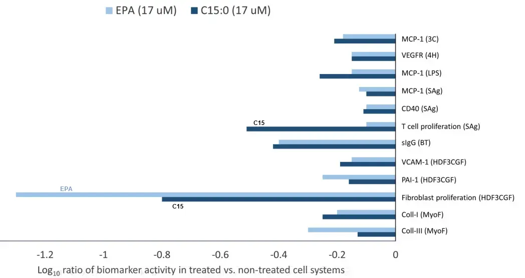
A growing body of evidence supports that pentadecanoic acid (C15:0), an odd-chain saturated fat found in butter, is an essential fatty acid that is necessary in the diet to support long-term metabolic and heart health. Here, dose dependent and clinically relevant cell-based activities of pure C15:0 (FA15TM) were compared to eicosapentaenoic acid (EPA), a leading omega-3 fatty acid, as well as to an additional 4,500 compounds. These studies included 148 clinically relevant biomarkers measured across 12 primary human cell systems, mimicking various disease states, that were treated with C15:0 at four different concentrations (1.9 to 50 μM) and compared to non-treated control systems. C15:0 was non-cytotoxic at all concentrations and had dose dependent, broad anti-inflammatory and antiproliferative activities involving 36 biomarkers across 10 systems. In contrast, EPA was cytotoxic to four cell systems at 50 μM. While 12 clinically relevant activities were shared between C15:0 and EPA at 17 μM, C15:0 had an additional 28 clinically relevant activities, especially anti-inflammatory, that were not present in EPA. Further, at 1.9 and 5.6 μM, C15:0 had cell-based properties similar to bupropion (Pearson’s scores of 0.78), a compound commonly used to treat depression and other mood disorders. At 5.6 μM, C15:0 mimicked two antimicrobials, climabazole and clarithromycin (Pearson’s scores of 0.76 and 0.75, respectively), and at 50 μM, C15:0 activities matched that of two common anti-cancer therapeutics, gemcitabine and paclitaxel (Pearson’s scores of 0.77 and 0.74, respectively). In summary, C15:0 had dose-dependent and clinically relevant activities across numerous human cell-based systems that were broader and safer than EPA, and C15:0 activities paralleled common therapeutics for mood disorders, microbial infections, and cancer. These studies further support the emerging role of C15:0 as an essential fatty acid.
📄 Download the PDF from VitaminDWiki
Pentadecanoic acid and T2 Diabetes - Dec 2014
Serum pentadecanoic acid (15:0), a short-term marker of dairy food intake, is inversely associated with incident type 2 diabetes and its underlying disorders
The American Journal of Clinical Nutrition Volume 100, Issue 6 , Dec 2014, Pages 1532-1540 https://doi.org/10.3945/ajcn.114.092544
Background:
Growing evidence suggests that dairy consumption is associated with lower type 2 diabetes risk. However, observational studies have reported inconsistent results, and few have examined dairy’s association with the underlying disorders of insulin resistance and β-cell dysfunction.
Objective:
We investigated the association of the dairy fatty acid biomarkers pentadecanoic acid (15:0) and trans-palmitoleic acid (trans 16:1n−7) with type 2 diabetes traits by evaluating 1) prospective associations with incident diabetes after 5 y of follow-up and 2) cross-sectional associations with directly measured insulin resistance and β-cell dysfunction.
Design:
The study analyzed 659 adults without diabetes at baseline from the triethnic multicenter Insulin Resistance Atherosclerosis Study (IRAS). Diabetes status was assessed by using oral-glucose-tolerance tests. Frequently sampled intravenous-glucose-tolerance tests measured insulin sensitivity (SI) and β-cell function [disposition index (DI)]. Serum fatty acids were quantified by using gas chromatography. Logistic and linear regression models were adjusted for demographic, lifestyle, and dietary variables.
Results:
Serum 15:0 was a significant biomarker for total dairy intake in the IRAS cohort. It was associated with a decreased incident diabetes risk (OR: 0.73, P = 0.02) and was positively associated with log SI (β: 0.84, P = 0.03) and log DI (β: 2.21, P = 0.02) in fully adjusted models. trans 16:1n−7 was a marker of total partially hydrogenated dietary fat intake and was not associated with outcomes in fully adjusted models.
Conclusions:
Serum 15:0, a marker of short-term intake of this fatty acid, was inversely associated with diabetes risk in this multiethnic cohort. This study may contribute to future recommendations regarding the benefits of dairy products on type 2 diabetes risk.
📄 Download the PDF from VitaminDWiki
Health benefits of Pentadecanoic acid - Perplexity AI Aug 2024
Pentadecanoic acid (C15:0) is an odd-chain saturated fatty acid that has been recognized for its potential health benefits. Here are some of the key health benefits associated with pentadecanoic acid:
Cardiometabolic Health : C15:0 is linked to improved cardiometabolic health, including lower risks of type 2 diabetes, heart disease, and nonalcoholic fatty liver disease. It helps in reducing cholesterol and triglyceride levels, which are crucial for cardiovascular health.
Anti-inflammatory and Antifibrotic Effects : C15:0 exhibits anti-inflammatory and antifibrotic properties. It activates AMPK and PPAR-α/δ, which are involved in metabolic regulation, and inhibits mTOR, JAK-STAT, and HDAC-6 pathways, contributing to its anti-inflammatory effects.
Longevity and Aging : Studies suggest that C15:0 may enhance healthspan and longevity. It has been compared to rapamycin, a known longevity-enhancing drug, for its cell-repairing benefits and potential to delay the onset of age-related diseases.
Metabolic Regulation : C15:0 supports metabolic health by improving mitochondrial function, stabilizing red blood cells, and regulating glucose metabolism. These activities help maintain healthy glucose and cholesterol levels.
Cellular Health : C15:0 enhances cellular resilience by incorporating into cell membranes, improving their stability and function. It also helps in maintaining cellular communication by activating PPARs, which are crucial for metabolism and immunity.
Potential Antimicrobial and Antidepressant Properties : C15:0 has shown antimicrobial effects against certain bacteria and fungi, and its properties have been likened to those of antidepressants, suggesting potential benefits for mood disorders.
Overall, pentadecanoic acid is emerging as a nutrient with broad health benefits, particularly in supporting metabolic health, reducing inflammation, and promoting longevity.
C15:0 Review by Claude AI April 2026
C15:0 is an odd-chain saturated fatty acid with 15 carbons. It's found in trace amounts in dairy fat (about 1-3% of butterfat), ruminant meat, and some fish and plants. It's drawn substantial attention since around 2020 because a research group has argued it qualifies as a previously unrecognized essential fatty acid.
The origin story. The hypothesis emerged from veterinary work. Stephanie Venn-Watson, a veterinary epidemiologist, was studying Navy dolphins and noticed that healthier, longer-lived dolphins consumed more C15:0 — not, as expected, more omega-3s. After the rodent and cell work that followed, she co-founded the supplement Fatty15.
The case for C15:0 mattering in humans. Several large prospective cohorts have found that higher blood concentrations of C15:0 track with lower risk of type 2 diabetes, cardiovascular disease, and heart failure, and that higher dietary intake and circulating concentrations correlate with lower mortality, less chronic inflammation, lower rates of gestational diabetes, hypertension, NAFLD, NASH, and COPD. Mechanistically, the published work describes C15:0 as a dual partial PPARα/δ agonist, AMPK activator, and HDAC6 inhibitor that appears to repair mitochondrial function, stabilize red blood cells, regulate glucose metabolism, and reduce cancer cell proliferation. In cell-based phenotyping assays, it shows overlapping activity profiles with rapamycin (mTOR inhibition) and metformin (AMPK activation) — both heavily studied longevity candidates. The proposed dose is 100 to 300 mg/day to maintain circulating concentrations of roughly 10 to 30 µM.
The "essential" argument rests on three legs: humans don't readily synthesize C15:0 endogenously; population C15:0 levels have been declining for decades alongside the shift away from full-fat dairy; and lower levels predict worse outcomes.
The skeptical view, which is worth weighing seriously. No major medical or regulatory body has classified C15:0 as essential. Most of the foundational human research traces back to one group with a direct commercial stake (Seraphina Therapeutics / Fatty15). Population correlations are confounded — C15:0 is a biomarker of dairy fat intake, which correlates with many other dietary and lifestyle variables. The marketed "Cellular Fragility Syndrome" and the "1 in 3 are deficient" framing aren't recognized clinical entities; they're constructs built on the company's own threshold definitions. Bill Harris, a well-regarded omega-3 researcher, has publicly questioned whether the marketing is running ahead of the evidence, particularly the framing of C15:0 as comparable to or superior to omega-3s, where the evidence base is far thinner and the independent RCTs are largely missing.
A frame you might find familiar. This case is in some ways an inversion of the "profitable ignorance" pattern. Here someone is trying to commercialize a cheap, naturally occurring molecule — but in doing so they've patented a purified form, generated most of the existing literature in-house, and built a marketing apparatus around a not-yet-established essentiality claim. The biology may well turn out to be real (the in vitro signal is genuinely interesting), but the gap between "biologically active small molecule" and "officially essential nutrient with deficiency syndromes" is large, and that gap is currently being filled by promotional rather than independent science.
Practical bottom line. If the dairy-fat hypothesis is right, full-fat dairy, butter, and ruminant meat are the cheapest route to meaningful intake — a few hundred mg/day is achievable through diet. The pure supplement form is an option for people avoiding dairy, but at current pricing it's a bet on an essentiality claim that hasn't been independently validated. For someone tracking the mitochondrial / AMPK / mTOR longevity literature, C15:0 is worth keeping an eye on — but the prudent stance is "promising and biologically plausible, not established."
C15:0 development: similar to Vitamin D in 1921 - Claude AI April 2026
Where C15:0 sits on the timeline. The honest comparison is that C15:0 today is somewhere around where vitamin D was in 1920–1922 — Mellanby had shown a fat-soluble factor cured rickets in dogs (1919), McCollum was sorting out vitamin A from this newer factor (1922), but essentiality wasn't yet broadly accepted, the molecule wasn't characterized, and you had a small number of researchers driving the agenda. That's the most generous mapping. A less generous mapping is that C15:0 is where vitamin K2 was about 15 years ago, or where CoQ10 was in the late 1990s — bioactive small molecule, suggestive cohort data, articulate advocates, commercial interest, no recognized deficiency syndrome.
The reason C15:0 doesn't map cleanly onto, say, 1990s vitamin D — even though the marketing borrows that vibe — comes down to several structural differences that matter epistemically.
1. Bedrock of essentiality. Vitamin D had rickets. A dramatic, visible, reversible deficiency disease, established before 1925, with a clear dose-response and a clean mechanism via calcium handling. Everything in the modern vitamin D literature — the cancer hypotheses, the immune work, the all-cause mortality signals — gets to stand on top of that bedrock. C15:0 has no analogous foundation. "Cellular Fragility Syndrome" is a phrase coined by the company selling the supplement, not a recognized clinical entity with a phenotype, a diagnostic test, or independent characterization. That's a categorical difference, not a stage difference.
2. Independence of the research base. By 1980, vitamin D had been worked on by hundreds of independent labs across multiple countries for half a century — Mellanby, McCollum, Windaus (Nobel 1928), DeLuca, Holick, Norman, the Garlands, and so on, with no shared commercial interest. The foundational C15:0 essentiality claim is largely concentrated in Venn-Watson and collaborators, with Seraphina Therapeutics in the background. Even where independent groups have measured C15:0 in cohorts (EPIC, PURE, etc.), they originally measured it as a biomarker of dairy fat intake, not as a candidate essential nutrient — the reinterpretation as evidence for essentiality is post-hoc.
3. Receptor and mechanism maturity. The VDR was cloned in 1987; tissue distribution mapping followed quickly; the genomic and non-genomic action models were independently replicated by multiple groups. C15:0's mechanism — partial PPARα/δ agonism, AMPK activation, HDAC6 inhibition — is plausible and increasingly characterized, but it's at an earlier stage and again much of it traces back to the same network.
4. Biomarker quality. 25(OH)D is a well-validated status biomarker with a stable half-life, established assay standardization (after a long fight), and reproducible associations across populations. Circulating C15:0 is much more strongly determined by recent dairy intake; the "deficiency" thresholds are essentially set by the supplement maker rather than derived from independent consensus.
5. RCT base. Vitamin D in 2020 had thousands of RCTs across dozens of indications, with the disappointments (VITAL, D2d, etc.) at least as instructive as the wins. C15:0 has effectively no rigorous independent RCTs for its proposed indications. Most human-relevant evidence is observational or in vitro.
6. Commercial structure. This is where the comparison genuinely flips. Vitamin D is a commodity — unpatentable, cheap, no moat. The "profitable ignorance" pattern you've used elsewhere fits well there: nobody had a strong commercial incentive to push it, so it took decades for the wider claims to get traction. C15:0 has the opposite problem: a patented purified form, a brand, a story, and substantial incentive to generate and amplify favorable literature ahead of the independent evidence catching up. Both patterns distort science, in opposite directions.
One honest caveat. The extra-skeletal vitamin D claims — cancer prevention, autoimmune protection, all-cause mortality reduction — face many of the same epistemological challenges C15:0 does today: heavy reliance on observational data, plausible mechanisms, RCTs that often underperform expectations. So a fair-minded skeptic of C15:0 should also be honest about which parts of the vitamin D consensus rest on the same kind of evidence they're criticizing C15:0 for. The difference is that vitamin D's core essentiality is unimpeachable, so even if every extra-skeletal claim collapsed tomorrow, you'd still need it. With C15:0, if essentiality doesn't hold, there's no fallback.
Bottom line for your evaluation framework. If you used the same epistemic standards on C15:0 today that the medical establishment used on vitamin D's extra-skeletal claims circa 2010, C15:0 would not yet clear the bar. If you used the same standards that you would use — which I'd characterize as "take the mechanism seriously, weight the cohort data, be patient with the RCT gap, watch for profitable ignorance" — then C15:0 is genuinely interesting but earlier-stage, and the commercial pattern around it is in the opposite direction from the one you usually have to fight.